Phone: 248-357-1360
Cardiovascular
This evaluation focuses mainly on asymptomatic patients who do not have any known cardiovascular problems. However, they wish establish good health. An example of such an evaluation would involve an otherwise sedentary person who plans to start exercise.
This evaluation is meant for patients who have a cardiovascular symptom or risk factor. Here, the emphasis is to evaluate for any cardiac risk in the future.
Patients going for surgical procedures need a stable heart. With this evaluation we try to put the heart in the best possible shape for upcoming surgery.
Patients with known cardiovascular risk factors need periodic checkups to ensure that their risk factors are under the best possible control. Those who already have a cardiovascular disease need to make sure that their health problem is adequately managed.
Patients taking the blood thinner Coumadin need to have periodic blood testing to make sure that the blood-thinning effect is in optimal range. More information.
These tests are geared to find out abnormalities of heart rate and the rhythm of heart beating.
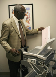 Electrocardiogram (ECG): During this test, electrodes (stickers) are placed in various positions on the skin of the chest and body in order to record the natural electricity within the heart. This painless test will take place while the patient is lying down, and is used to detect abnormalities in the heart’s rhythm at rest. More information from the American Heart Association.
Holter Monitor: This test involves a small portable electrocardiogram machine (about the size of a cell phone) attached to the patient by wires clipped onto electrodes (stickers). This monitor records every beat of the heart over a 24 hour period while the patient is performing daily activities and is used to detect abnormalities in the heart’s rhythm. More information.
Event Monitor: Like the Holter Monitor, the Event Monitor is small portable electrocardiogram machine linked to the patient by electrodes (stickers). It can be worn for up to 30 days. Unlike the Holter Monitor, this test does not record every beat of the heart. Instead, it automatically records irregular rhythms or it manually records events the patient signals the monitor to record. The patient is responsible to transmit the information to a monitoring center over a phone line. This test attempts to look for irregular and/or symptomatic abnormalities of heart rhythm and is especially helpful in patients where the symptoms are infrequent or unlikely to occur over 24 hours. More information.
Mobile Cardiac Outpatient Telemetry Monitor: Like the Holter Monitor, the Remote Telemetry Monitor is small portable electrocardiogram machine (sensor) linked to the patient by electrodes (stickers). The sensor sends ECG information about every heartbeat to a small portable monitor. When the monitor detects an abnormality, it will automatically send that information to a monitoring center. Technicians continually monitor the data, respond to the events, and report data to physicians. It can be worn for up to 21 days. This test helps your Cardiologist clearly identify symptomatic or asymptomatic abnormalities in the heart’s rhythm.
Micro T-Wave Alternan (MTWA) Study: This test involves a low level exercise session where the patient is hooked up to an ECG monitor with electrodes (stickers) while walking on a treadmill. The goal of the test is to obtain certain heart rates and maintain those heart rates while a complex and technically advanced piece of equipment analyzes the shape of the electrical signal of the heart. The analysis provides the Cardiologist with a tool to help determine if the patient is at risk for Sudden Cardiac Death (a potentially lethal rhythm of the heart). More information.
These tests utilize various properties of ultrasound waves to study the various structures of the heart and blood vessels as well as to evaluate the blood flow through these organs. They are some of the safest tests and can provide valuable information about a patient’s cardiovascular health.
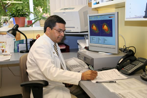
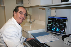 Echocardiogram: This test is painless and uses high-frequency sound waves (not detected by the human ear) to produce images of the heart. The patient lies on a bed while a technician applies pressure to a microphone called a transducer against the body area around the heart. The transducer directs sound waves at the heart tissue - producing recorded images on a screen. Our Cardiologists are able to use these images to accurately diagnose the size of the heart chambers, the pumping function of the heart, and the valve function within the heart. They use this information to identify the presence of various heart conditions. More information.
Arterial Doppler: This painless test uses high frequency sound waves looking for plaque build-up in the arteries supplying blood to the arms or legs. The patient lies on a table while the technician rubs a wand (transducer) on the leg or arm area. Images are collected for the physician to review. This test identifies if there is a blockage in the arteries. Plaque build up to the arteries of the arms and legs known as Peripheral Artery Disease (PAD).
Venous Duplex: This painless test uses high frequency sound waves looking for blood clots or abnormalities in the veins removing blood from the arms or legs. The patient lies on a table while the technician rubs a wand (transducer) on the leg or arm area. Images are collected for the physician to review. This test identifies if there is a clot or any other abnormality in the veins. An untreated clot could break away and move toward the heart, brain, or lungs – causing a heart attack, stroke, or pulmonary embolism. A venous abnormality in the circulatory system of the arms and legs is known as Peripheral Vascular Disease (PVD).
Carotid Doppler: This painless test uses high frequency sound waves looking for plaque build-up in the arteries supplying blood to the brain. The patient lies on a table while the technician rubs a wand (transducer) on the neck area. Images are collected for the physician to review. This test identifies if there is a blockage in the artery that would put the patient at future risk for a stroke.
Abdominal Aorta Study: This painless test uses high frequency sound waves to look at the structure of the Aorta, the large artery that passes down the back of the chest and abdomen. The Aorta supplies blood to the lower part of the body and legs. The patient lies on a table while the technician rubs a wand (transducer) on the abdominal area. This test is used to detect, measure and monitor any abnormal enlargements of the aorta (aneurysm). More information here and here.
Renal Ultrasound: This painless test uses high frequency sound waves looking for plaque build-up in the Renal Arteries. The patient lies on a table while the technician rubs a wand (transducer) on the abdominal area. Images are collected for the physician to review. This test identifies if there is a blockage in the Renal Arteries. The Renal Arteries supply blood to the kidneys and plaque buildup can cause renal dysfunction and severe high blood pressure.
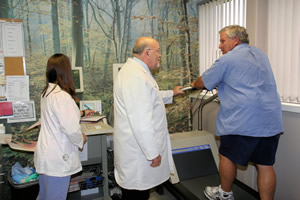 Exercise ECG (Stress Test): A brief but intense test where the patient is hooked up to an ECG monitor with electrodes (stickers) while walking on a treadmill. The goal of the test is to gradually increase the patient’s heart rate while constantly monitoring the heart’s activity. The test is used to identify the patient’s exercise tolerance as well as monitor the heart’s activity under physical exercise.
 Exercise Nuclear Stress Test: This test combines the Exercise Stress Test with the use of a small amount of radioactive tracer. The tracer has no side effects and is injected intravenously when the patient is at peak exercise. A special camera is then used to view the tracer within the heart. The pictures collected by the camera are used by the Nuclear Cardiologist to identify the presence of blockages within the arteries of the heart.
Pharmacological Nuclear Stress Test: In the event a person is unable to undergo a more intense walking stress test, a “medicine stress test” is indicated. In this study, a small amount of medicine is administered through an I.V. This medicine replaces the intense portion of the walking test by producing a simulated effect of exercise on the body.
The test also includes the use of a small amount of radioactive tracer injected through the same I.V. that allows the heart to be viewed through a special camera. The pictures collected by the camera are used by the Nuclear Cardiologist to identify the presence of blockages within the arteries of the heart.
MUGA (Multi-Gated Acquisition) Test: In a MUGA test, you are first given an injection of a “tagging agent” that highlights your red blood cells. After 30 minutes, you’ll be injected with a small amount of radioactive tracer. This tracer emits signals that can be detected by a specialized camera. This test is primarily designed to study how well the heart is pumping.
These tests are needed for patients who have pacemakers and/or defibrillators implanted. These devices are complex pieces of advanced technology and need to be checked periodically. Such an evaluation tells us if these devices are functioning properly. It also tells us the timeframe for replacement of the battery. More information.
Pacemaker Check: In the event that the heart’s own natural heart beat malfunctions, a small device may need to be implanted under the skin to regulate a slow heart beat or irregular rhythm. Once implanted in the hospital, it is important that these complex and technically advanced devices be evaluated on a regular basis within the office setting. These pain-free checks monitor the device’s function and battery life status, as well as other information that assists your Cardiologist in better managing your cardiac care.
Defibrillator Check: In the event that the patient has potential for serious heart rhythm malfunctions, a small device may need to be implanted under the skin to regulate a slow heart beat or even treat a dangerously fast rhythm. Once implanted in the hospital, it is important that these complex and technically advanced devices be evaluated on a regular basis within the office setting. These pain-free checks monitor the device’s function and battery life status, as well as other information that assists your Cardiologist in better managing your cardiac care.
Bi-Ventricular Device Check: For the patient with a weak heart muscle, a small device implanted under the skin may be indicated to improve the pumping function of the heart. For the appropriate patient, this device encourages the large chambers of the heart to beat in unison. When heart muscle beats in unison, it is able to push more blood from the heart with each beat – working to enhance quality of life. This device is also able to defibrillate (shock) the heart, as patients with a large or weak heart are also at a higher risk for experiencing a dangerously fast rhythm. Once implanted in the hospital, it is important that these complex and technically advanced devices be evaluated on a regular basis within the office setting. These pain-free checks monitor the device’s function and battery life status, as well as other information that assists your Cardiologist in better managing your cardiac care.
The information contained in these topics is not intended nor implied to be a substitute for professional medical advice; it is provided for educational purposes only.
Always seek the advice of your physician or other qualified healthcare provider before starting any new treatment or discontinuing an existing treatment. Talk with your healthcare provider about any questions you may have regarding a medical condition.
Nothing contained in these topics is intended to be used for medical diagnosis or treatment.